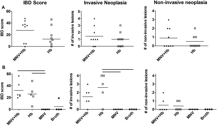
Figure 4.
IBD and neoplasia in Smad3−/− mice infected with H. bilis alone or coinfected with MNV and H. bilis. IBD scores (left), number of invasive lesions (middle), and number of noninvasive lesions (right) are shown for (A) study 2 and (B) study 3. In study 3, 2 additional control groups of mice were infected with MNV alone (n = 6) or broth only (n = 4) are shown. Black bars above graphs in (B) indicate statistically significant differences between groups as tested by nonparametric one-way ANOVA and a Dunn post test. There were no statistically significant differences between mice coinfected with both MNV and H. bilis or with H. bilis only (P > 0.05 for Mann–Whitney tests of IBD and tumor scores; P > 0.05 for Fisher exact test for tumor incidence).

