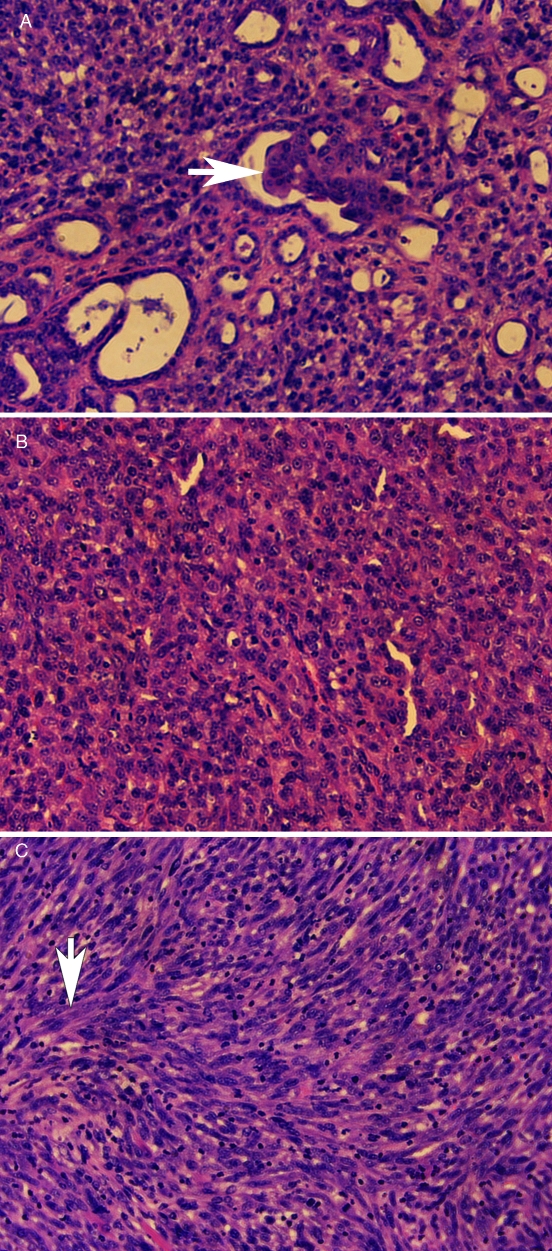
Figure 3.
Histopathology of an MDCK cell tumor in an adult nude mouse. (A) An epithelial portion of tumor with irregular tubule formation, some with papillary ingrowths (arrow). (B) Tumor with polygonal cells. (C) Tumor with fusiform cells arranged in interwoven bundles (arrow). The tumors were fixed in formalin, paraffin-embedded, sectioned, stained with hematoxylin and eosin, and photographed under a 10× objective.

