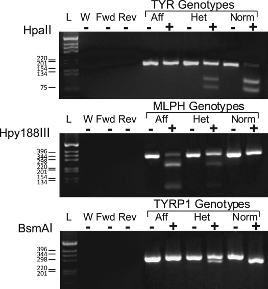
Figure 2.
Scheme of the TYR, MLPH, and TYRP1 PCR–RFLP diagnostics. The images are of the electrophoresis gels of the PCR amplicons and resultant fragments. Control PCR reactions included: complete reaction mixtures minus DNA template (W), complete reactions with only the forward (Fwd) or reverse (Rev) primer, and control DNA samples from known affected (Aff), carrier (Het), and normal (Norm) cats. For each representative genotyped, there are 2 lanes: 1 containing the uncut PCR product (–) and the other the PCR product cut with the enzyme indicated at the left of the illustration (+). On the left is the DNA ladder with ladder marker sizes indicated.

