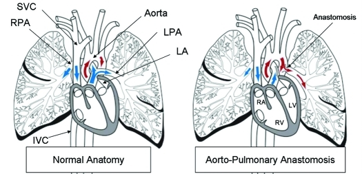
Figure 1.
Schematic of normal anatomy and the aortopulmonary anastomosis. At surgery, the left pulmonary artery (LPA) was connected to the descending aorta. IVC, inferior vena cava; LA, left atrium; LV, left ventricle; RA, right ventricle; RPA, right pulmonary artery; RV, right ventricle; SVC, superior vena cava.

