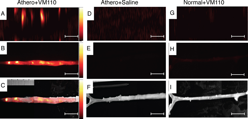
Figure 3.
In vivo high-resolution NIRF molecular imaging of cysteine proteinase activity in atherosclerosis. (A) Aorta from atherosclerotic rabbits injected with Prosense VM110 one day prior, and imaged in vivo with the intravascular NIRF catheter. (B) Ex vivo FRI at 800 nm and (C) ex vivo NIRF-white light fusion image. (D) Aortas from atherosclerotic rabbits injected with saline and imaged in vivo with the NIRF catheter, and (E) ex vivo FRI at 800nm and (F) ex vivo NIRF-white light fusion image. (G) Normal rabbit injected with VM110 and imaged with the intravascular NIRF catheter. (H) Ex vivo FRI at 800nm (1 sec) and (I) ex vivo NIRF-white light fusion image. In vivo and ex vivo fluorescence images were equally windowed and processed (color lookup table applies to all figures in each row). Scale bar, 10 mm.

