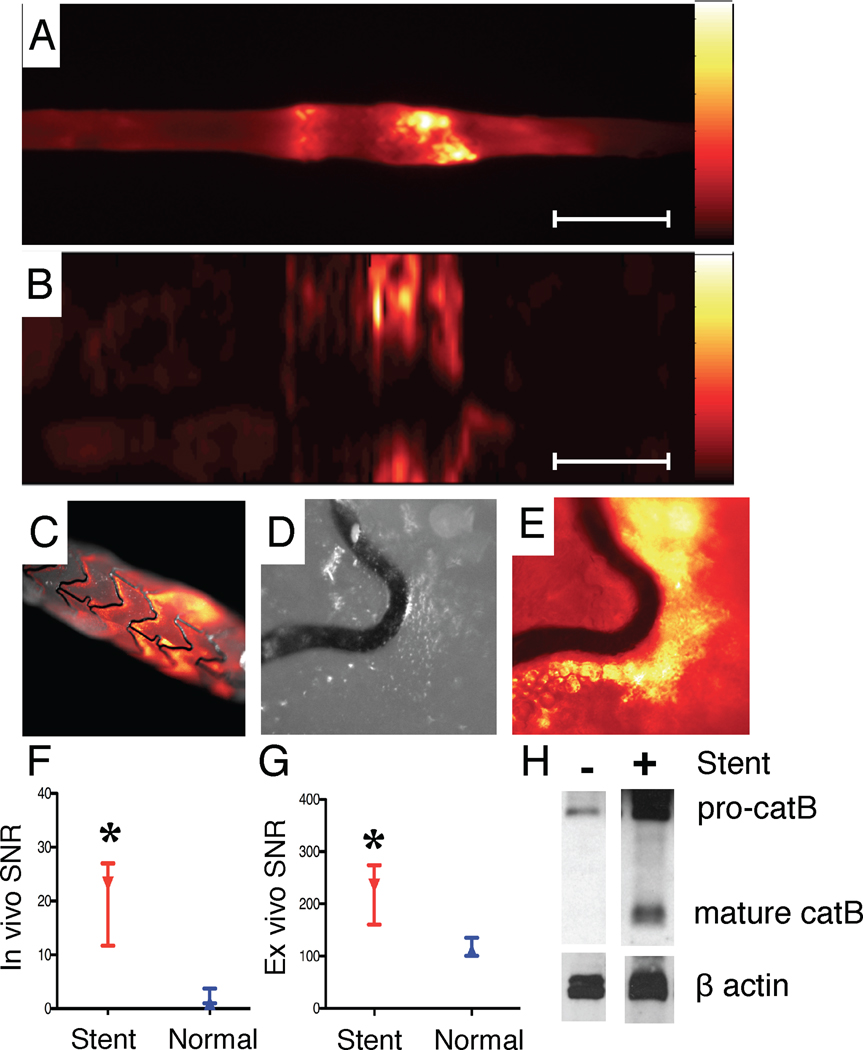
Figure 9.
NIRF imaging of stent injury. (A) Ex vivo FRI at 800nm reveals augmented NIRF protease activity at stent edges. (B) Corresponding ex vivo intravascular NIRF pullback also detected stent-based NIRF signal increases. (C) Ex vivo NIRF-white light fusion image of the stent, (D) high-magnification white light and (E) high-magnification NIRF image reveals signal along the greater curvature of stent struts. (F) In vivo and (G) ex vivo SNR in the normal and stented aorta (paired observations). (H) Immunoblot of cathepsin B (catB) staining in normal and stented rabbit aorta. Pro-catB denotes the 46 kD pre-cathepsin B band, and mature catB denotes the 25 kDa and 30 kDa cathepsin B bands. Scale bar, 10 mm. *p<0.05.

