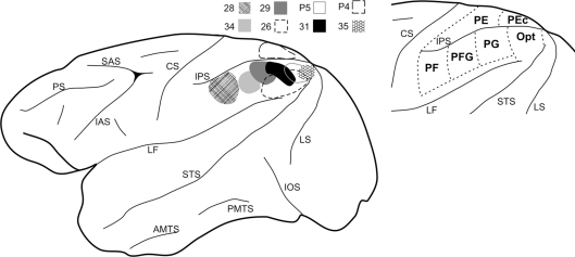
Figure 1.
Summary of the parietal injection sites used for this study. At left, the BDA injections (n = 8) are schematically mapped onto a lateral view of a macaque monkey left hemisphere. A schematic mapping of parietal lobe subdivisions is shown at the right, as adapted from Pandya and Seltzer (1982) and Gregoriou et al. (2006). AMTS, anterior middle temporal sulcus; CS, central sulcus; IAS, inferior arcuate sulcus; IOS, inferior occipital sulcus; IPS, intraparietal sulcus; LF, lateral fissure; LS, lunate sulcus; PMTS, posterior middle temporal sulcus; PS, principal sulcus; SAS, superior arcuate sulcus; STS, superior temporal sulcus.

