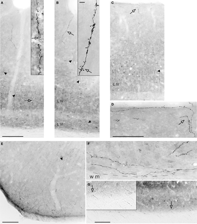
Figure 3.
Photomicrographs of axons in the CF, anterogradely labeled by BDA injections in IPL. (A,B) Two sequential sections from ventral V1 (see Figure 4C). Labeled segments are visible in all layers (arrowheads). Higher magnification (insets) show terminations in layer IV (A) and I, II (B), where the hollow arrows point to corresponding features at the two magnifications. (C) Another field of BDA labeling in V1. Terminations in layer I (hollow arrow) are shown at higher magnification in (D). (E) A field of BDA-labeled terminations in dorsal V2. Arrowhead indicates an ascending axon segment. (F) BDA-labeled terminations in layer VI of area V1. The dashed line indicates white matter border (WM). (G) A field of labeled axon segments in the white matter subjacent to V1. Inset, from hollow arrow, shows five segments at higher magnification. (E) is from Case 28, all the other examples are from Case P5. Scale bar in (A) applies to (B) and (C) (200 μm); scale bar in the inset in (B) applies to inset in (A) (10 μm); scale bar in (D), applies to (F) and inset in (G) (100 μm). Scale bars in (E) and (G) = 100 μm.

