
Figure 3.
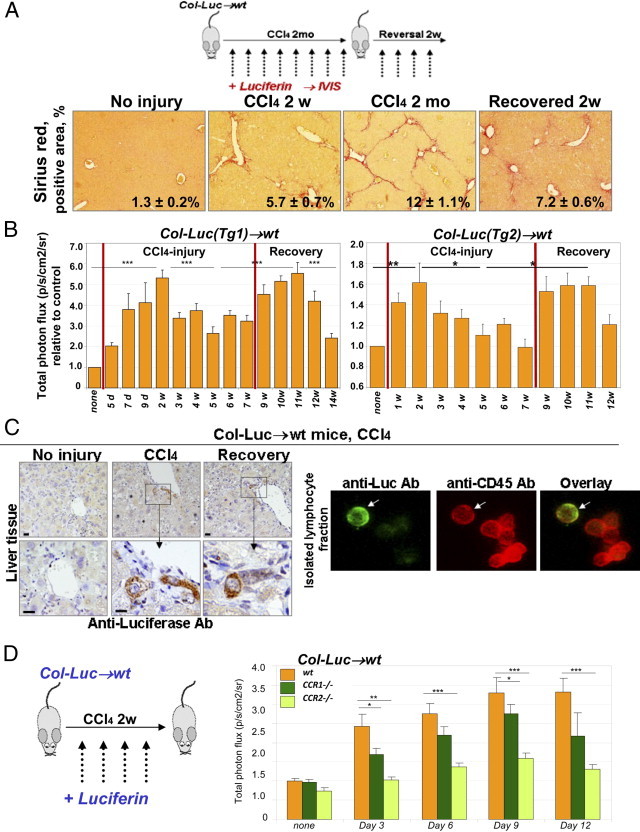
CCR2 and CCR1 facilitate fibrocyte recruitment in response to CCl4. A: Study design (top). Col-Luc→wt mice were treated with CCl4 and recovered from fibrosis. Sirius red staining (bottom) determined the total collagen deposition at chosen times (mean ± SEM; P < 0.05). B: CCl4-treated Col-Luc→wt mice were monitored using bioluminescence. Luc+ cells egress BM in two phases corresponding to acute injury and recovery (n = 10). *P < 0.05, **P < 0.01, and ***P < 0.001. Groups compared are none and 2w, 2w and 5w, 5w and 11w, and 11w and 14w for Col-Luc(Tg1)-Luc mice. Graphic representation of the biphastic egress of fibrocytes and precursors in response to CCl4. C: Livers and fibrocyte-containing fraction4 from Col-Luc→wt mice are stained with anti-Luc and anti-CD45 antibodies. Scale bars = 50 μm. D: Recruitment of Luc+ cells was studied in LucCCR2−/−→wt mice (n = 10) and LucCCR1−/−→wt mice (n = 10) mice and compared with Lucwt-into-wt mice in response to CCl4 (2 weeks). Bars represent mean ± SEM. *P < 0.05, **P < 0.01, and ***P < 0.001. Groups compared are wt and LucCCR2−/−→wt mice, and wt and LucCCR1−/−→wt mice.