
Figure 4.
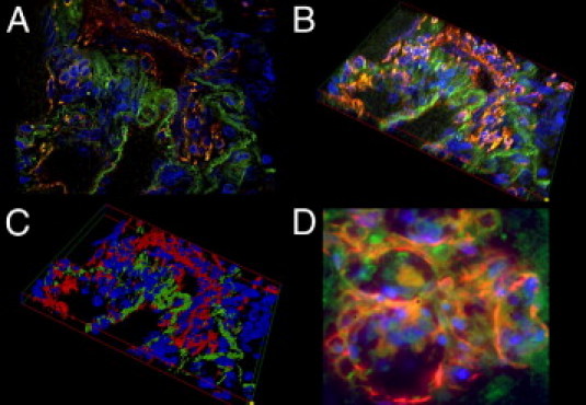
The-3-D reconstruction of a PL. A: A fluorescent double-stained PL: the luminal vascular channels stain positive for CD31 (red) and the adjacent interstitium shows homogenous positivity for SMA (green). B: Systematically scanned images of the whole width of the PL were merged into 3-D shapes. C: Images were skeletonized into a voxel model for increased clarity. Note the distinct separation of the endothelial layer and the interstitium in the PL. D: GLLs in high-grade neural tumors show a rather disorganized composition without an equally clear separation of endothelial layer and interstitium. Original magnification: ×630 (A–C); ×1000 (D).