
Figure 1.
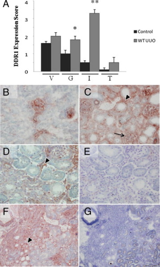
DDR1 expression is induced in the interstitium of the obstructed kidney. A: Semiquantitative evaluation of DDR1 expression in the kidney 12 days after UUO in vessels (V), glomeruli (G), interstitium (I), and tubular epithelium (T). B–G: DDR1 immunostaining in the control kidney. B: In control kidney, predominant expression is in the arteriolar wall and in the mesangium (×40). Then 12 days after UUO, DDR1 is strongly induced in interstitial cells (arrowheads) (C and D, ×40; F, ×20). Note a focal de novo expression on glomerular epithelial cells (arrow). E: Negative control (×40). F and G: Proximal tubular epithelial cells, identified by megalin-positive apical staining, do not significantly coexpress DDR1, as evidenced by DDR1 (F) and megalin (G) stainings on consecutive sections (×20). Data are mean ± SEM. *P < 0.05, *P < 0.01 versus nonobstructed WT kidney.