
Figure 2.
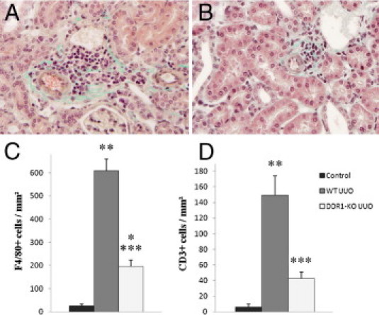
DDR1−/− mice present reduced accumulation of inflammatory cells in the obstructed kidney. Representative views of Masson trichrome stainings in obstructed kidneys in WT (A, ×40) and DDR1−/− mice (B, ×40). Note the cellular infiltration in the vicinity of the vessels. Cortical cell count for F4/80+ cells (C) and CD3+ cells (D) in control, WT UUO, and DDR1−/− UUO kidneys, expressed per unit area. Data are mean ± SEM. *P < 0.05, **P < 0.01 versus nonobstructed kidney; ***P < 0.01 versus obstructed WT kidney.