
Figure 1.
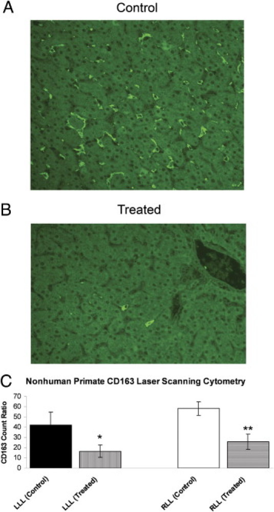
Nonhuman primate (cynomolgus macaque) liver section CD163 and KCs by laser-scanning cytometery (LSC). A: Liver section from a control monkey showing many scattered KCs. B: Liver section from a drug-treated monkey (the drug is a novel neutralizing human M-CSF monoclonal antibody) showing significantly fewer KCs. C: LSC quantitation. LLL indicates left lateral lobe; and RLL, right lateral lobe. There was a significant decrease in KCs in treated animals. *P < 0.05, **P < 0.01.