
Figure 12.
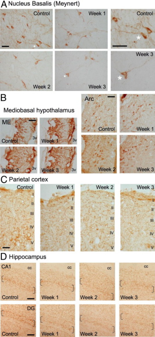
Time-course effects of Aβ25-35 (10 μg/rat) intracerebroventricular injection on VAChT immunolabeling within the nucleus basalis of Meynert (A), mediobasal hypothalamus (B), parietal cortex with levels I to V cortical layers indicated (C), and hippocampus (D) were determined in control untreated rats and at 1, 2, and 3 weeks after Aβ25-35 injection. In A, asterisks indicate matching locations in corresponding images presented at both lower and higher magnification. In D, brackets locate the hippocampus granular cell layers. Arc, arcuate nucleus; cc, corpus callosum; ME, median eminence; 3v, third ventricle. Scale bars = 100 μm.