
Figure 5.
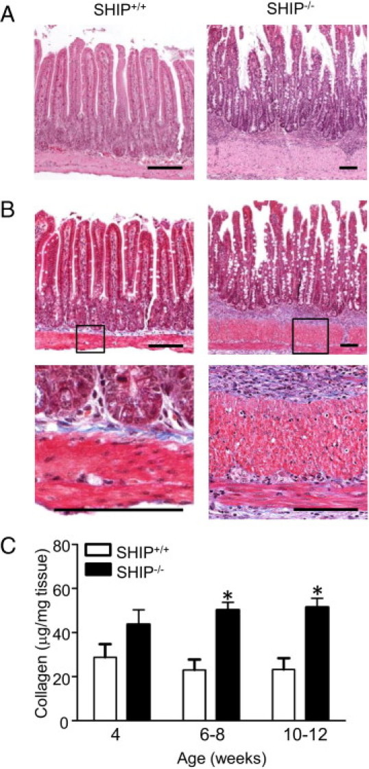
SHIP–/– mice have elevated collagen deposition in their distal ilea, indicative of fibrosis. A: H&E-stained horizontal sections of the distal ileum of 8-week-old SHIP+/+ (left) and SHIP–/– (right) mice revealed a thickened muscle layer in SHIP–/– mice. B: Masson's trichrome stain (blue) of adjacent sections from the same mice revealed increased collagen deposition. Insets: Higher magnifications show muscularis externa and muscularis mucosa and the presence of collagen between muscle layers (bottom). Scale bars = 100 μm. C: Sircol assays comparing collagen levels between SHIP+/+ and SHIP–/– ilea. Bars represent the mean ± SD for 6 to 12 mice at each age (*P < 0.05).