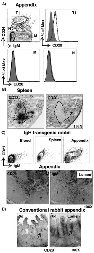
Figure 3. Tissue localization of CD20+ transitional B cells.

A) Flow cytometric staining of appendix B cells from a neonatal rabbit (1 wk of age) for T1 (CD24hiIgMlo) (dashed), M (CD24-IgM+) (black) and non-B cells (N) (IgM-CD24-) (black) for CD20. Shaded histogram = isotype control. B) Immunohistological staining of spleen (6-week old) section for CD23 (follicular B cells) and CD20 (transitional B cells). The dotted line represents a B cell follicle. C) Flow cytometric and immunohistological analyses of tissues from an IgH transgenic rabbit stained for IgM and CD21 (upper), and CD20 and IgM (lower), respectively. D) Staining for CD20 in appendix from a conventional 3 & 6-day-old rabbit. Magnification = 100X.
