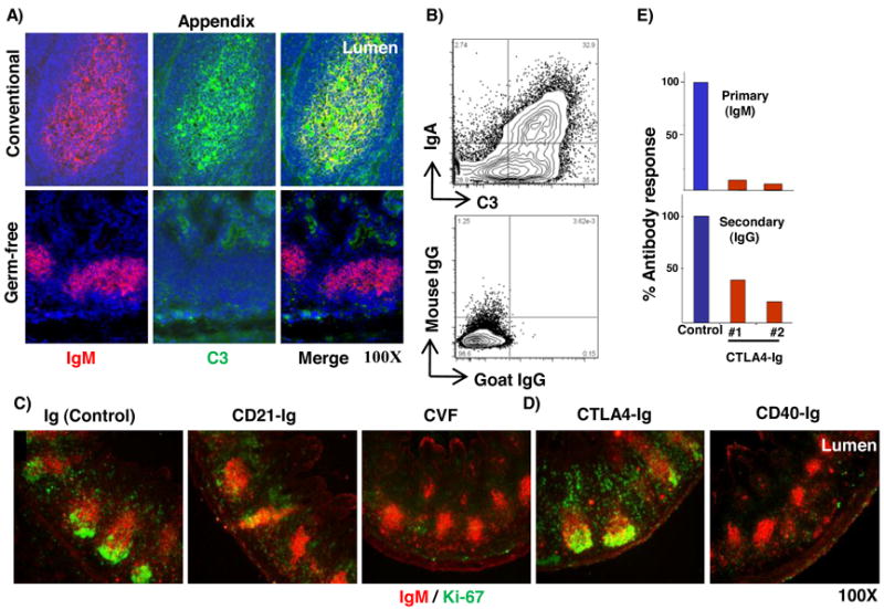
Figure 5. Identification of molecules required for proliferative expansion of B cells in GALT.

A) Immunofluorescent staining for IgM and C3 in appendix sections from conventional 4 wk old rabbit (upper) and 4 wk old rabbit with a germ-free appendix (lower). B) Flow cytometric analysis of IgA- and C3- stained intestinal commensal bacteria from 4 wk old rabbit. Plots are representative of two independent experiments. C) & D) Immunofluorescent staining of appendix sections for IgM and Ki-67 following treatment of newborn rabbits with adenovirus (Ad) expressing soluble receptors; Ig (negative control), CD21 (CR2-Ig), cobra venom factor (CVF), CTLA4 (CTLA4-Ig) and CD40 (CD40-Ig). Data are representative of 3 or more Ad- or CVF-treated rabbits. E) Bar graph showing the primary anti-BGG (IgM) (upper) and secondary anti-BGG (IgG) (lower) response, compared to the anti-BGG response from an age-matched littermate control (=100%) as determined by ELISA. #1 and #2 represent data from two rAdCTLA4-Ig treated rabbits. Magnification = 100X.
