
Figure 4.
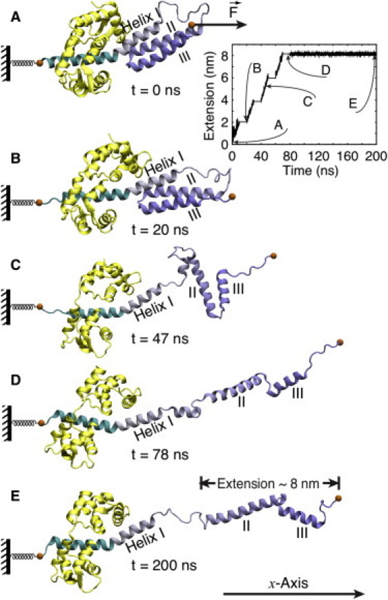
Stretching of myosin VI PT domain. Shown are snapshots during the force-induced extension process at (A) t = 0 ns; (B) t = 20 ns; (C) t = 47 ns; (D) t = 78 ns; and (E) t = 200 ns. Extension is found to proceed in two steps: a first step (A → B → C) involves dissociation of helices II and III from helix I; a second step (C → D) involves loss of contact between helix II and helix III. (Inset) Sequence of pulling and relaxation intervals during the 200-ns simulation (see text). The zig-zag extension process and the subsequent relaxation is shown in Movie S1 in the Supporting Material.