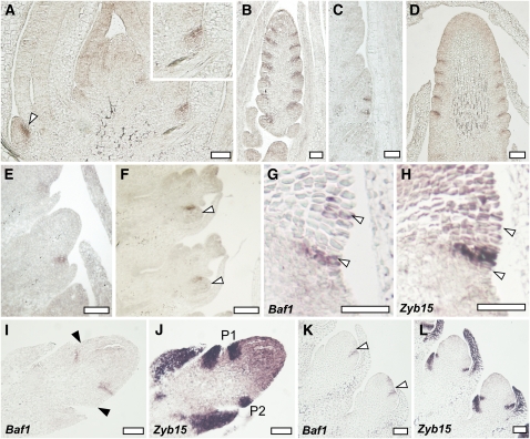
Figure 6.
The Expression Pattern of Baf1.
(A) to (F) In situ hybridizations of Baf1 during vegetative and reproductive development in the wild type.
(A) Longitudinal section of a seedling showing Baf1 expression adaxial to the newly developing axillary meristems (arrowhead and close-up).
(B) and (C) Localization of Baf1 in an immature tassel (B) and a tassel branch (C) in a narrow domain where the axillary meristems are forming.
(D) A similar localization is observed in immature ears.
(E) and (F) In developing spikelet meristems, Baf1 is localized where a new floral meristem is forming (arrowheads).
(G) to (L) Consecutive sections hybridized with probes for either Baf1 or Zyb15, a marker for suppressed bract primordia.
(G) and (H) flank of an inflorescence meristem. Arrowheads point to a few Baf1-expressing cells in (G) and the corresponding region in (H).
(I) and (J) Immature developing ear.
(K) and (L) Developing spikelet meristems.
Black arrowheads in (I) mark the corresponding P1 and P2 sites of Zyb15 expression in (J). White arrowheads in (K) mark the Baf1 expression domain. P1 and P2, suppressed bract primordia. Bars = 50 μm.

