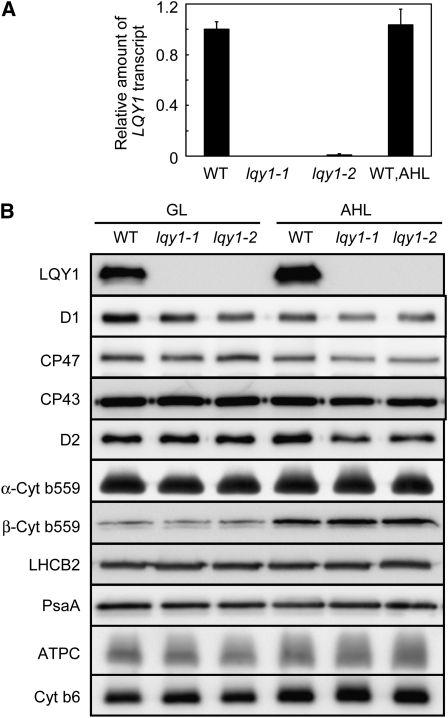
Figure 4.
Analysis of LQY1 Transcript and Thylakoid Membrane Protein Accumulation.
(A) Relative amount of the LQY1 transcript determined by quantitative RT-PCR. The amount of LQY1 transcript was normalized by that of the ACT2 transcript (At3g18780). Values (mean ± se, n = 3) are given as the ratio to the amount of LQY1 transcript in wild type (WT) under growth light. For LQY1 transcript levels after a 2-d high-light treatment, only the wild-type value is shown because the transcript was not detectable in either lqy1 mutant. AHL, after 2 d of high light.
(B) Representative immunoblots of LQY1 and PSII proteins in wild-type and lqy1 mutants. Thylakoid membrane proteins were separated by SDS-urea-PAGE, electroblotted to a PVDF membrane, and probed with affinity-purified anti-LQY1 or antisera against known thylakoid membrane proteins obtained from Eva-Mari Aro or Agrisera Co. The lanes on each gel were loaded on an equal chlorophyll basis. Cyt, cytochrome; GL, growth light.

