
Figure 1.
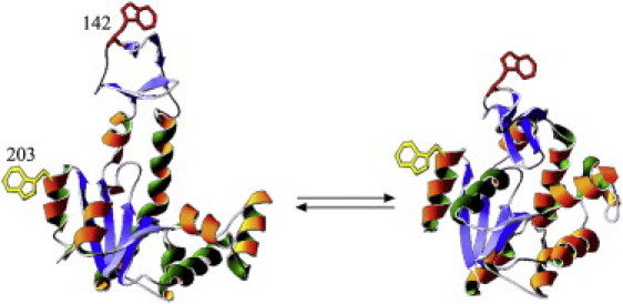
Crystal structures of E. coli AK in the ligand free state (PDB 4AKE) (left) and when bound to the two substrate mimicking inhibitor Ap5A (PDB 1AKE). The labeled sites are indicated both in 203 and in 142 by an indole side chain.
Official websites use .gov
A
.gov website belongs to an official
government organization in the United States.
Secure .gov websites use HTTPS
A lock (
) or https:// means you've safely
connected to the .gov website. Share sensitive
information only on official, secure websites.
Crystal structures of E. coli AK in the ligand free state (PDB 4AKE) (left) and when bound to the two substrate mimicking inhibitor Ap5A (PDB 1AKE). The labeled sites are indicated both in 203 and in 142 by an indole side chain.