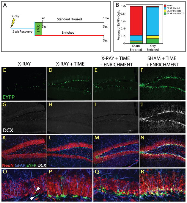
Figure 6. X-irradiation obliterates neurogenesis but not stem cell accumulation.
(A) Animals were subjected to sham or X-irradiation, allowed 2 weeks recovery, exposed to standard or enriched (EEE) housing, and treated with TMX. Animals were sacrificed immediately after TMX (standard housed only) or after 1 month of standard housing or EEE. (B) The proportion of cellular populations within the EYFP+ lineage in a representative sham and x-irradiated EEE animal. The majority of EYFP+ cells are neurons in sham-, and NSCs in X-irradiated animals. Confocal micrographs reveal the lack of DCX after X- (G–I) but not sham irradiation (J), indicating that DCX is not expressed in the irradiated brain. (C, K, O) X-irradiation does not obliterate all EYFP+ cells. (O) Surviving EYFP+ cells are GFAP+ and exhibit mainly NSC morphology. Some stellate GFAP+EYFP+ cells are also present. There are more EYFP+ cells in X-irradiated animals 1 month after TMX (D,L,P) and most cells are GFAP+ NSCs (P). The number of EYFP+ cells is similar in EEE (E, M) and standard housed (D, L) X-irradiated animals. (P, Q) Radial processes are more tortuous in NSCs of EEE X-irradiated animals than EEE shams. Higher magnification reveals that most EYFP+ cells are GFAP+ and display radial morphology in X-irradiated (O–Q), but not sham (R) animals.

