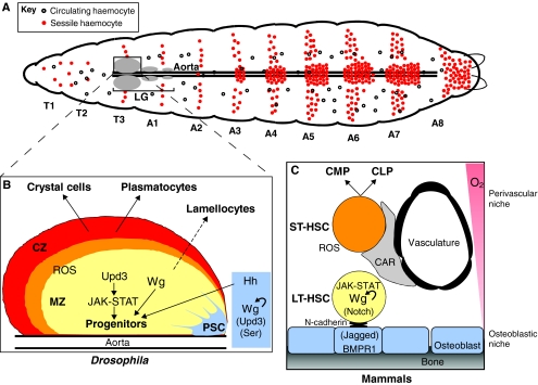
Fig. 1.
Parallels in haematopoiesis between the Drosophila LG and mammalian bone marrow. (A) Schematic representation of a Drosophila third instar larva, illustrating the presence of circulating and sessile haemocytes of embryonic origin, and showing the position of the larval haematopoietic organ (the LG) along the dorsal aorta. T and A refer to thoracic and abdominal segments, respectively, and are numbered along the anterior-posterior axis. (B) The primary lobe of the LG consists of the PSC (blue), the MZ (yellow; containing haemocyte progenitors) and the CZ (red), in which differentiation of plasmatocytes and crystal cells takes place. Lamellocytes only differentiate in response to specific immune threats, at the expense of progenitors. There also exists a population of intermediate progenitors that have left the MZ (found within the orange region). Signalling pathways that are active in the PSC and MZ are indicated. Tightly controlled levels of ROS sensitise haemocyte progenitors to differentiation. Unpaired 3 (Upd3) is one activator of the JAK-STAT pathway, and Serrate (Ser) and Jagged are ligands of the Notch receptor. Their specific roles in the niche remain little understood. (C) Schematic representation of the quiescent LT-HSCs and primed (activated) ST-HSCs and their niches in mammalian bone marrow. Signalling pathways involved in the communication between HSCs and their niches are indicated. Common lymphoid and common myeloid precursors are indicated as CLP and CMP, respectively. Curved arrows indicate cell-autonomous effects within the PSC (B) or in LT-HTCs (C).

