Fig. 2.
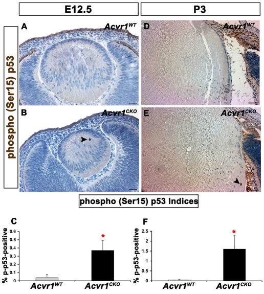
Acvr1CKO lenses have increased phosphorylation of p53 at Ser15. Acvr1WT and Acvr1CKO lenses were stained for phosphorylated (p)-p53. (A) A representative image of an E12.5 Acvr1WT lens with no detectable p-p53 staining. (B) p-p53-positive fiber cell nuclei were detected in Acvr1CKO lenses at E12.5 (arrowhead). (C) The percentage of p-p53-positive fiber cells was significantly higher in Acvr1CKO lenses than in Acvr1WT lenses at E12.5. (D) A representative Acvr1WT lens with no detectable p-p53-positive cells at P3. (E) An Acvr1CKO lens with a p-p53-positive fiber cell nucleus at P3 (arrowhead). (F) At P3, significantly more p-p53-positive fiber cells were detected in Acvr1CKO lenses than in Acvr1WT lenses. *P<0.05. Scale bars: 20 μm (A,B), 50 μm (D,E).

