Fig. 6.
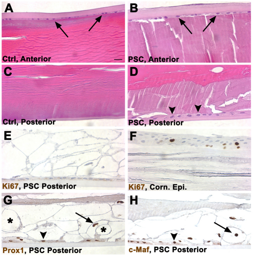
The appearance of cells in a human PSC is similar to those in Trp53CKO mouse lenses. (A) The anterior surface of a normal adult human lens is bounded by the thick anterior capsule and a thin layer of lens epithelial cells (arrows). Beneath the epithelium are layers of well-organized superficial fiber cells. (B) The anterior surface of an adult human lens with a PSC appears similar to the normal lens in A. (C) The posterior surface of the normal lens has a thinner capsule than does the anterior of the lens. Beneath the capsule are layers of superficial fiber cells. (D) The posterior of the lens with a PSC has a layer of globular ‘balloon’ cells between the capsule and the well-organized, deeper fiber cells. A layer of epithelioid cells is present on the inner surface of the capsule (arrowheads). (E) No Ki67-labeled nuclei were detected in balloon or epithelioid cells at the posterior of the lens that had PSCs. The epitope unmasking method used in the staining procedure extracted the cytoplasm from the ‘balloon’ cells, causing them to appear as ‘ghosts’. No Ki67-positive nuclei were detected in the anterior epithelium from this lens (not shown). (F) Several nuclei in the corneal epithelium from the eye with a PSC were Ki67 positive, indicating that the staining procedure worked as expected. (G) The nuclei of balloon (arrow) and epithelioid (arrowhead) cells in the PSC stained with an antibody to the fiber-cell-specific marker Prox1. Some of the empty ‘ghosts’ resulting from the epitope unmasking method are labeled with an asterisk. (H) The nuclei of balloon (arrow) and epithelioid (arrowhead) cells in the PSC stained with an antibody to the fiber-cell-specific marker Maf.

