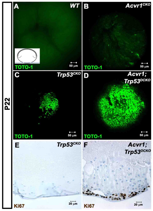
Fig. 7.
Loss of Acvr1 leads to proliferation in posterior subcapsular plaques, forming ‘lens tumors’. P22 lenses were stained with the nucleic acid stain TOTO-1 and their posterior poles were examined using a confocal microscope. The inset in A shows the plane of section and region of the lens that was viewed. (A) No nuclei were seen in the posterior poles of Acvr1WT lenses. Staining of RNA in the fiber cell cytoplasm shows the posterior lens sutures. (B) A few scattered nuclei were evident near the posterior surface of Acvr1CKO lenses. (C) A small accumulation of nuclei was present at the posterior pole of Trp53CKO lenses. (D) A large mass of cells was present at the posterior poles of Acvr1;Trp53DCKO lenses. (E) Antibody to Ki67 showed that cells in the posterior subcapsular plaques of Trp53CKO lenses were not in the cell cycle. (F) Antibody to Ki67 showed that cells in the posterior subcapsular plaques of Acvr1;Trp53DCKO lenses were still in the cell cycle.

