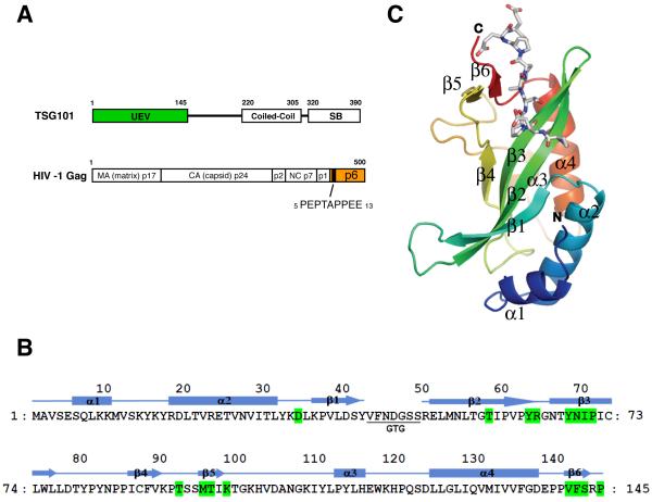
Figure 1. Structure of TSG101 UEV and HIV-1 p6 peptide.
(A) Schematic representations of human TSG101 and HIV-1 Gag. Abbreviations within TSG101 are: UEV (ubiquitin E2 variant) and SB (‘steadiness’ box). Viral protease cleavage sites are indicated by vertical lines and the names of resulting proteins are shown. The location of the PTAP peptide in p6Gag is indicated. (B) Amino acid sequence and secondary structure of TSB101 UEV. The residues involved in PTAP peptide binding are colored in green. (C) Overall structure of TSG101 UEV and HIV p6 peptide.

