
Figure 4.
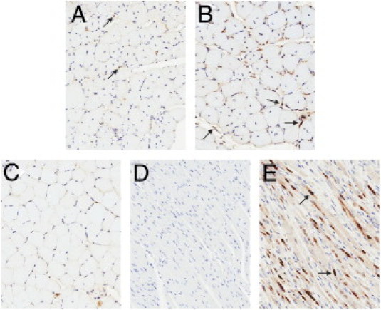
Myogenin immunostained skeletal muscle. Representative digital scans of myogenin immunostained soleus muscle sections from mdx mice treated from 2 weeks to 9 months of age with vehicle (A), 1D11 (B), or vehicle-treated wild-type mice (C), along with negative controls (rat heart, D) and positive controls (rat heart injected with rat skeletal muscle myocytes, E). Arrows indicate myogenin positive nuclei. Original magnification, ×20.