
Figure 6.
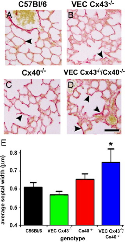
Collagen deposition in lungs from VEC Cx43−/−/Cx40−/− mice. Paraffin-embedded distal lung sections from C57Bl/6 (A), VEC Cx43−/− (B), Cx40−/− (C), and VEC Cx43−/−/Cx40−/− (D) mice at age 32 weeks were labeled using Picro-sirius Red to stain collagen, and were imaged. Compared with the other strains of mice examined, lungs from VEC Cx43−/−/Cx40−/− mice showed enhanced collagen staining (arrowheads). Scale bars = 60 μm. E: Paraffin-embedded distal lung sections from C57Bl/6, VEC Cx43−/−, Cx40−/−, and VEC Cx43−/−/Cx40−/− mice at age 32 weeks were analyzed using morphometric analysis. Compared with the other strains examined, VEC Cx43−/−/Cx40−/− mice demonstrated significantly thicker alveolar septal width between the pulmonary endothelium and the alveolar epithelium. *P < 0.05.