
Figure 5.
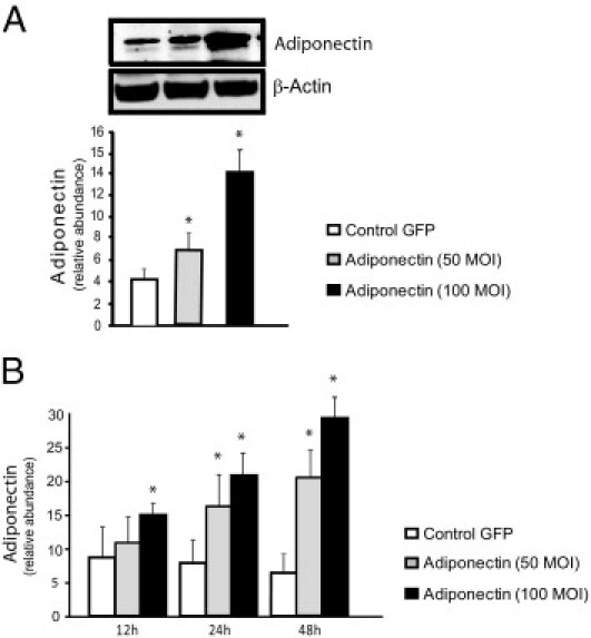
Quantification of adiponectin protein expression in hepatic stellate cells. Stellate cells from WT mice on FVB background were isolated, cultured for up to 48 hours, and then infected with a lentivirus expressing GFP, or adiponectin at 50 and 100 MOI. Cell lysates were harvested at 48 hours (A) or at 12, 24, and 48 hours (B) and were subjected to immunoblotting. A representative immunoblot is shown (A). Subsequently, specific bands from repeated experiments were quantified and normalized to the signal for β-actin (A and B) (means ± SE; n = 3). *P < 0.05 versus control GFP.