
Figure 4.
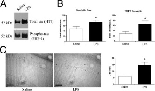
Insoluble tau and phospho-tau is increased in LPS-treated 3xTg-AD mice. A: Representative HT7 and PHF-1 immunoblot of formic acid fractions shows increased levels of tau and phosphorylated tau in LPS-treated mice. B: Quantification of HT7 and PHF-1 immunoblot shows significantly increased levels of tau and PHF-1–positive tau in the formic acid fraction of LPS-treated mice (*P < 0.05, n = 5). C: Representative PHF-1 reactivity shows increased levels of PHF-1 in LPS-treated mice. PHF-1–immunoreactive neurons in the subiculum and CA1 hippocampus are significantly increased in LPS-treated mice (*P < 0.05, n = 5). Scale bar = 100 μm.