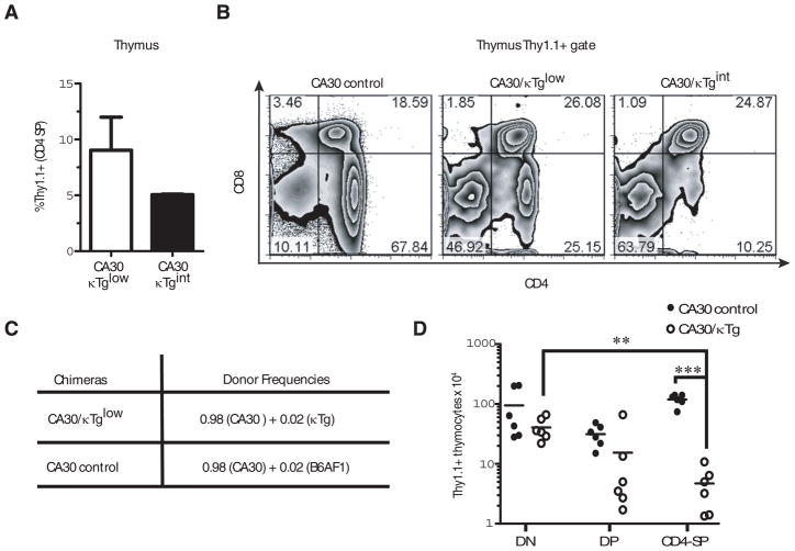
Figure 3. Partial deletion of CA30 thymocytes in chimeras with κTg B cells.
A, Mean percent of total CD4+ SP thymocytes that were Thy1.1+ in CA30/κTg mixed BM chimeras. B, Distribution of CA30 (Thy1.1+) thymocyte subpopulations in representative CA30/κTg chimeras. Thymocytes were segregated on the basis of the Thy1.1 marker (CA30 cells), and analyzed for the expression of CD4 and CD8. Error bars indicate SEM (n=5). C, Donor BM ratios used to reconstitute lethally irradiated B6AF1 mice. D, Numbers of thymocytes in mixed chimeras generated as indicated in C (red blood cell lysed). Statistics were calculated using a one-tailed paired t test. Data represent the composite of two independent experiments. DN (CD4− CD8− double negative), DP (CD4+ CD8+ double positive) and CD4 SP (single positive).** p=0.0023 *** p< 0.0001.

