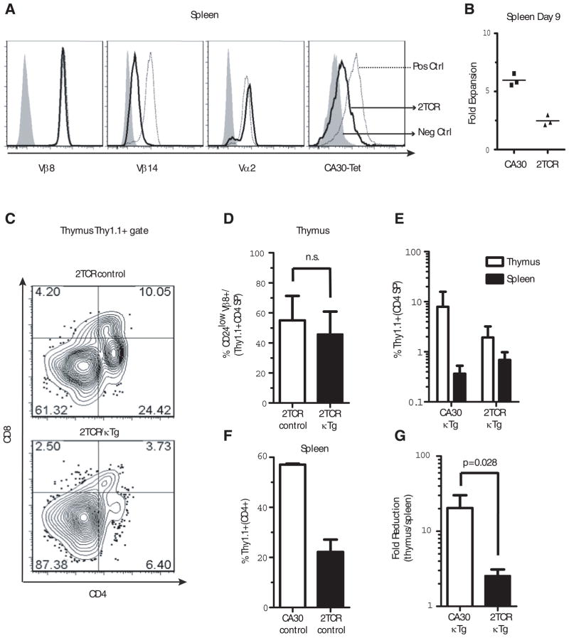
Figure 6. Expression of a second TCR rescues mature CA30 CD4 SP thymocytes.
A, Expression of various αβTCR chains by CA30/F508 2TCR T cells (dark line), positive controls (dotted line, CA30 1st and 4th panels, F508 2nd and 3rd panels) and negative controls (shaded histogram, F508 1st and 4th panels, CA30 2nd and 3rd panels). One of 15 mice. B, Diminished primary immune response by 2TCR T cells. 106 total lymph node cells (2TCR or CA30) were transferred into B6AF1 recipients that were immunized i.p. 1 day later with Vκ36–71 FR1 peptide (10 μg) + LPS (7 μg) or with PBS. At day 9, Thy1.1+ CD4+ cells were quantified and are shown as fold-increase in peptide immunized mice versus PBS-immunized mice. One of 2 experiments. C, Frequency of 2TCR CD4 SP thymocytes in representative experimental (2TCR/κTg) and control (2TCR/B6AF1) chimeras. D, Mean frequencies of mature 2TCR CD4 SP thymocytes in mixed chimeras containing κTg B cells (~6% engraftment) or B6AF1 B cells (control). E, Percent CA30 T cells among CD4+ cells in the thymus and spleen of BM chimeras. F, Poor engraftment of 2TCR cells relative to CA30 T cells. Both sets of chimeras made with B6AF1 BM. Percent of Thy1.1+ T cells in spleens of CA30 or 2TCR chimeric control mice (reconstitution ratio CA30 or 2TCR/B6AF1 80:20). G, Fold-reduction from thymus to periphery in 2TCR CD4+ and CA30 CD4+ cells in chimeras with κTg B cells. Values calculated using frequencies of total CD4+ T cells in periphery or total CD4 SP thymocytes that were Thy1.1+. Columns are mean plus SEM (panels C–G, n=4, n.s. = not significant).

