
Figure 3.
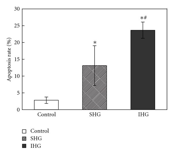
Detection of apoptotic cells with flow cytometry. After the treatment for 72 h, cells were collected for the determination of cell apoptosis as described previously. The percentage of apoptotic cells was shown after analysis by flow cytometry. Data were expressed as mean ± SD of three determinations. *P < .05 versus control, ∗# P < .05 versus SHG.