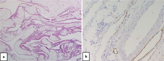
Fig. 3.
Histological findings. a Hematoxylin and eosin staining showed that the multicystic lesion was composed of an irregularly dilated space separated by fibrous tissue and smooth muscle fascicle. The wall of the cyst was lined by a layer of lymphatic endothelial cells. Those endothelial cells showed partially positive staining for D2-40 (b).

