
Figure 1.
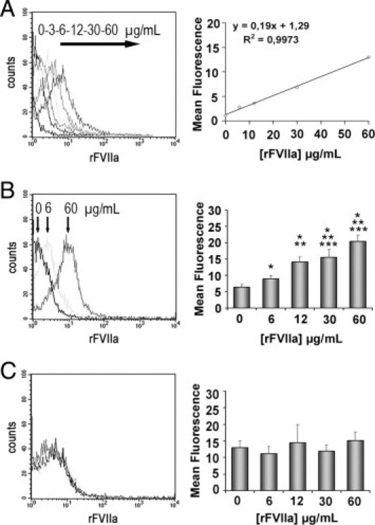
Flow cytometry detection of rFVIIa in platelets from healthy donors exposed to different doses of rFVIIa (0, 3, 6, 12, 30, and 60 μg/mL) for 2 hours. The leftpanels show the representative shift in the fluorescent profiles for the different concentrations of rFVIIa associated to the platelet population. A: Internalization of Alexa Fluor 488-labeled rFVIIa by platelets. The rightpanel shows the linear regression corresponding to the mean of fluorescence and the concentrations of AF488-rFVIIa used. B: Intraplatelet unlabeled rFVIIa detected through a two-step antibody method combined with a permeabilization procedure of the platelet membrane. The rightpanel shows a dose-dependent increase of intraplatelet mean fluorescence measured for each concentration of unlabeled rFVIIa tested. C: No statistical differences were observed in the mean of fluorescence corresponding to rFVIIa associated to the platelet membrane. *P < 0.01 versus PBS; **P < 0.05 versus rFVIIa 6 μg/mL; and ***P < 0.05 versus rFVIIa 12 μg/mL (n = 10).