
Figure 3.
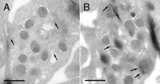
Immunostaining of FVIIa in platelets cryosections obtained from washed platelet suspensions previously incubated or not with rFVIIa 6 μg/mL for 2 hours. A: Immunoelectron micrograph showing background labeling for FVIIa (10-nm gold particles, arrows) on platelets that had not been incubated with rFVIIa. B: Micrograph corresponding to platelets incubated with rFVIIa. Positive labeling was detected for FVIIa in the membranes of the open canalicular system, in the platelet cytoplasm, and in the α granules. Labeling was also observed associated to the platelet surface after 2 hours of exposure to rFVIIa. Scale bars = 1000 nm.