
Figure 5.
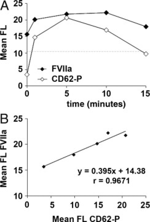
Exposure of FVIIa on platelet membrane after platelet activation with thrombin. Washed platelets previously incubated with 30 μg/mL of rFVIIa were activated with thrombin (0.1 U/mL) for 0, 1, 5, 10, and 15 minutes. A: Sequential flow cytometry analysis showing exposure of FVIIa (solid symbols) and CD62-P (open symbols) on the platelet membrane at the different time points. Results are expressed as mean fluorescence (FL). The dashed horizontal line indicates levels of mean fluorescence for FVIIa in platelets not exposed to rFVIIa. Platelet activation with thrombin resulted in the exposure of FVIIa on the platelet surface, which paralleled expression of CD62-P. B: Correlation studies between the mean of fluorescence measurements of CD62-P and FVIIa confirmed a good correspondence between the exposure of both antigens.