
Figure 1.
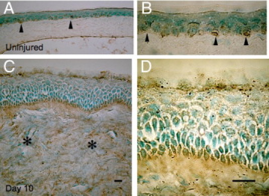
TRPV1-immunostaining in corneas of WT mice. The localization of TRPV1 in the intact mouse cornea is restricted to the basal cell layers of the epithelial cells (A, arrowheads). B: Higher magnification of the epithelium (arrowheads). On the other hand, TRPV1 is detected in stromal cells (asterisks) in a healing burned cornea besides basal epithelial cells (C). D: Higher magnification of the epithelium. Scale bar = 10 μm.