
Figure 2.
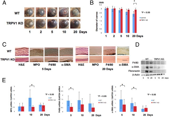
Healing of alkali-burned corneas in TPV1 KO mice. A: The corneas of WT and KO mice at 1, 2, 5, 10, or 20 days. On each of these days, the incidence and degree of opacification in the burned cornea was more prominent in WT mice than TRPV1 KO mice. At day 20, the iris is observed through the transparent cornea in a KO mouse, although not in a WT mouse. B: Evaluation of the alteration of the diameter of the eyeball during wound healing after alkali burn shows that WT globes have a smaller diameter at 20 days than the KO globes. C: Histology of burned corneas stained with H&E and IHC findings at days 5 and 20. H&E staining shows that the burned cornea shows a higher cell population and more severely disorganized tissue in WT corneas as compared with KO tissues at both days 5 and 20. The stroma was thicker in WT corneas as compared with KO corneas throughout the healing interval examined. IHC suggests that the density of the MPO-labeled cells at day 5 and F4/80-labeled cells at day 20 is greater in a WT cornea as compared with a KO cornea. Burned corneas that are healing seem to contain many α-SMA–positive myofibroblasts in WT mice at days 5 and 20. However, the majority of corneal fibroblasts were not labeled with anti–α-SMA antibody in TRPV1 KO mice. Scale bar = 100 μm. D: Western blotting shows expression of F4/80, α-SMA, and fibronectin were higher in WT tissue at 10 and 20 days after alkali burn. E: qRT-PCR also shows that the loss of TRPV1 suppresses mRNA expression levels of MPO, F4/80, and α-SMA during wound healing after alkali burn. Data represent mean ± SEM from five specimens in each condition. *P < 0.05.