
Figure 8.
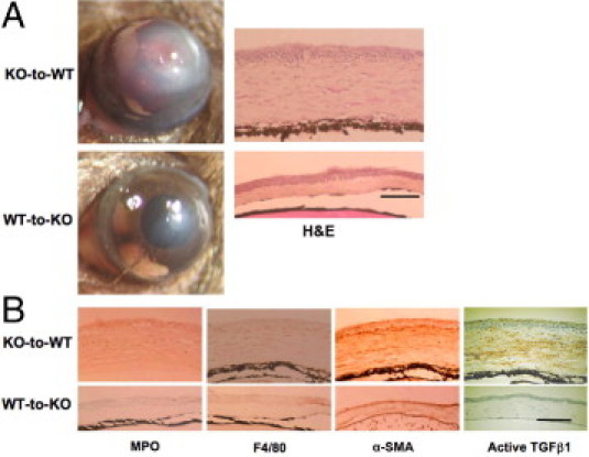
Healing an alkali-burned cornea in a mouse that has received a BMT at day 10 of healing. A: Ten days after alkali burning, a WT mouse that had received BM from a KO mouse (KO-to-WT group) showed much more opacification and neovascularization as compared with a KO mouse that had received BM from a WT mouse (WT-to-KO group). H&E histology shows larger cell populations in the swollen stroma of a KO-to-WT cornea as compared with WT-to-KO tissue. B: IHC shows that the cornea of a WT-to-KO mouse has less stromal α-SMA (the myofibroblast marker) staining as well as lower levels of immunoreactivity of MPO (a neutrophil marker), F4/80 (a macrophage marker), and active TGFβ1 as compared with the KO-to-WT tissue. Scale bar = 100 μm.