
Figure 9.
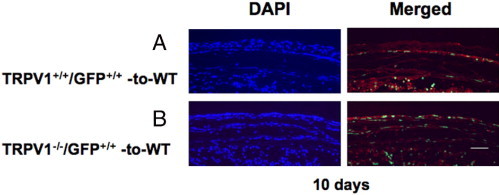
Healing an alkali-burned cornea in a mouse that has received GFP-positive BMT at day 10 of healing. A: Ten days after alkali burning, a WT mouse that had received BM from a TRPV1+/+/GFP+/+ mouse (TRPV1+/+/GFP+/+-to-WT group). B: WT mouse that had received BM from a TRPV1−/GFP+ mouse (TRPV1−/−/GFP+/+-to-WT group). Green staining shows GFP-positive cells. Red staining shows F4/80 localization and blue DAPI staining shows cell nuclei. Scale bar = 100 μm.