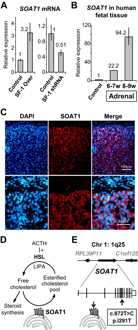
Fig. 2.
A, Increased and decreased levels of SOAT1 mRNA 48 h after SF-1 overexpression or knockdown, respectively, were confirmed by qRT-PCR [data represented according to the 2−ΔΔCT method (5)]. B, Expression of SOAT1 mRNA in human fetal adrenal tissue was assessed by qRT-PCR. Results showed a marked increase in SOAT1 expression from 6–7 to 8–9 wpc in comparison to control tissue (heart, 8 wpc), reflecting active steroidogenesis in the fetal adrenal cortex. C, Immunofluorescence confirmed strong expression of SOAT1 in the outer layers of the human fetal adrenal cortex at 8 wpc but not in overlying nonsteroidogenic capsule cells (DAPI, 4′,6-diamidino-2-phenylindol, was used to visualize nuclei). The omission of primary antibody resulted in no signal (data not shown). D, Cartoon representation of the actions of SOAT1 (HSL, hormone-sensitive lipase; LIPA, lipase A). E, Cartoon representation of the SOAT1 locus at 1q25 and of the allelic variant identified.

