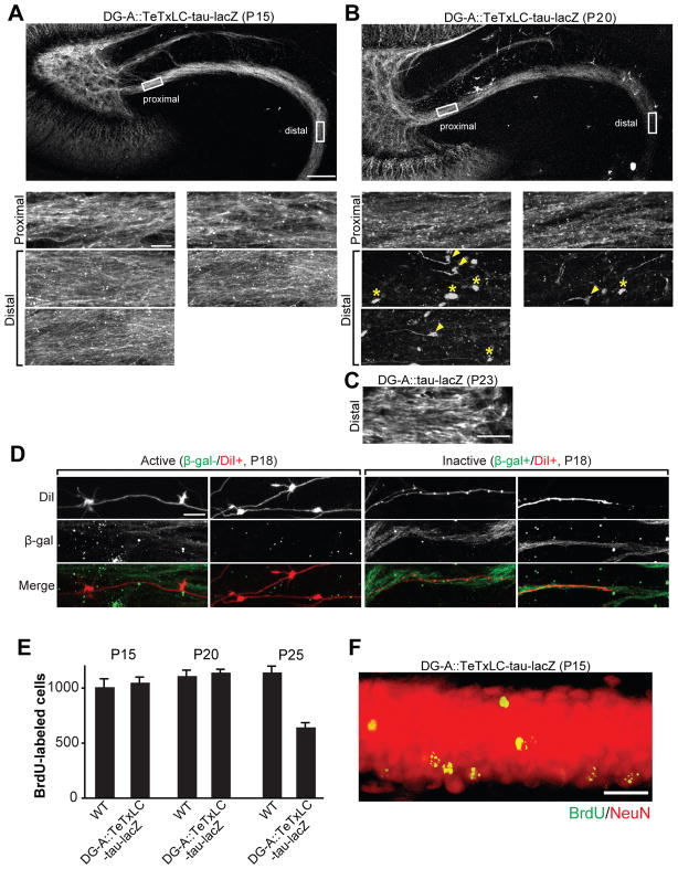
Figure 4. Elimination of Inactive DG Axons Is by Axon Retraction and Not Caused by DG Cell Death.
(A and B) Representative confocal images of TeTxLC-expressing DG axons in the DG-A::TeTxLC-tau-lacZ line at P15 (A) and P20 (B). 50 μm thick horizontal vibratome sections were stained with the anti-β-gal antibody to visualize TeTxLC-tau-lacZ-expressing axons. Higher magnification pictures of the proximal and distal regions of the axons are shown in the bottom panels. At P20 (B), at the distal region of DG axons, there were retraction bulbs (arrowheads) and remnants of axons (axosomes; asterisks), which are signs of axon retraction. Scale bars are 100 μm (top), 10 μm (bottom panels).
(C) Confocal image of the distal region of DG axons in the DG-A::tau-lacZ line (no TeTxLC) at P23 (anti-β-gal staining). Without TeTxLC, the distal region of DG axons remained intact at P23. Scale bar is 10 μm.
(D) Confocal images of DiI-labeled active and inactive DG axons in DG-A::TeTxLC-tau-lacZ mice. Hippocampal sections (100 μm thick) were prepared from P18 DG-A::TeTxLC-tau-lacZ mice and their DG axons were sparsely labeled with DiI. The sections were stained for β-gal to identify inactive axons. Active axons (β-gal-/DiI+) had many large mossy fiber buttons, whereas no large buttons were found in inactive axons (β-gal+/DiI+). Scale bar is 10μm.
(E and F) Wild type (WT) and DG-A::TeTxLC-tau-lacZ mice (15 mice each) received a single injection of BrdU (300 mg/kg) at P7–8 to label dividing DG cells. Hippocampal sections were prepared at P15, P20 and P25 (5 mice per day for each genotype) and stained for BrdU to evaluate DG cell survival. (E) The number of BrdU-positive DG cells at P15, P20 and P25. Similar numbers of BrdU-labeled DG neurons were found in WT and DG-A::TeTxLC-tau-lacZ mice at P20 when inactive DG axons were being retracted (B), suggesting that inactive DG axon retraction was not caused by DG cell death. The number of BrdU-positive cells in DG-A::TeTxLC-tau-lacZ mice was significantly decreased at P25, suggesting that inactive DG neurons were eventually eliminated after axon retraction. Bars are mean ± SEM. (F) Double staining for BrdU and NeuN of a DG section from DG-A::TeTxLC-tau-lacZ mice (P15). All BrdU-labeled DG neurons were NeuN positive. Scale bar is 20μm.
See also Figure S2.

