Fig. 2.
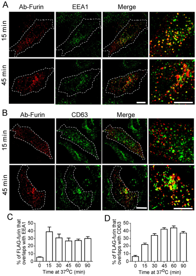
Furin is transported via the early and late endosomes en route to the TGN. HeLa cells were transfected with FLAG–furin for 24 hours and monolayers were incubated with anti-FLAG antibodies for 45 minutes on ice. Cells were washed in PBS and incubated in serum-free medium for 15 or 45 minutes at 37°C, then fixed, permeabilised and stained for internalised antibody–FLAG–furin complexes with Alexa-Fluor-568-conjugated anti-rabbit IgG (red) and for (A) EEA1 using mouse monoclonal anti-EEA1 antibodies (green) or (B) CD63 using mouse monoclonal anti-CD63 antibodies (green). Scale bars: 10 μm. The number of FLAG–furin pixels that overlapped with (C) EEA1 or (D) CD63 was expressed as a percentage (±s.e.m.) of the total number of FLAG–furin pixels within each cell using the plug-in OBCOL on the ImageJ program (n=20 for each time point).

