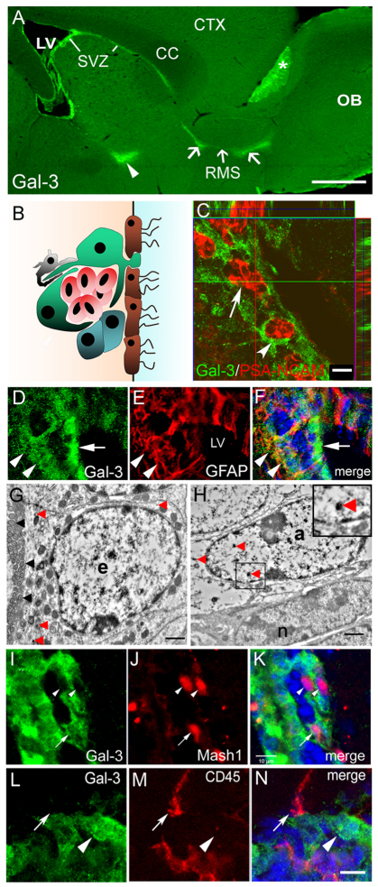
Fig. 1.
Gal-3 expression on astrocyte lineage cells surrounds neuroblast chains. (A) Expression of Gal-3 in the SVZ, along the RMS (arrows) and in the accessory olfactory bulb (asterisk). Arrowhead indicates a portion of the ventral SVZ that was included in the sagittal section. CC, corpus callosum; CTX, cerebral cortex; LV, lateral ventricle; OB, olfactory bulb. Scale bar: 250 μm. (B) Cytoarchitecture of the SVZ. Astrocyte-like stem cells (‘B cells’, green) and transit-amplifying progenitor cells (‘C cells’, blue) surround migratory neuroblasts (‘A cells’, red). Microglia (grey) interdigitate cells and ciliated ependymal cells (brown) line the ventricle. (C) Confocal image shows Gal-3 (arrowhead) is not expressed by PSA-NCAM+ neuroblasts (arrow), but Gal-3+ cells surround neuroblasts. Scale bar: 10 μm. (D–F) Gal-3 immunofluorescence is diffuse and is associated with some GFAP+ cells (arrowheads). Many ependymal cells also express Gal-3 (arrow). Blue in F is DAPI nuclear label. (G) Electron microscopy shows Gal-3 immunoreactivity (red arrowheads) in ependymal cell (e). Note the row of cilia (black arrowheads) on the left. Scale bar: 1 μm. (H) Gal-3 immunoprecipitates were observed in astrocytes (a) (arrowheads) but not in neuroblasts (n) using immunoelectron microscopy. Inset shows high magnification view of a Gal-3 immunoprecipitate. Scale bar: 1 μm. (I–K) Gal-3 (green) is expressed by few Mash1+ cells (arrow) in the SVZ. Arrowheads indicate Mash1+ cells that are Gal-3−. Scale bar: 10 μm. (L–N) Gal-3 is rarely expressed by CD45+ cells (red). Arrow shows close juxtaposition but lack of co-labeling. Arrowhead indicates a Gal-3+ cell that is CD45−. Scale bar: 10 μm.

