Fig. 2.
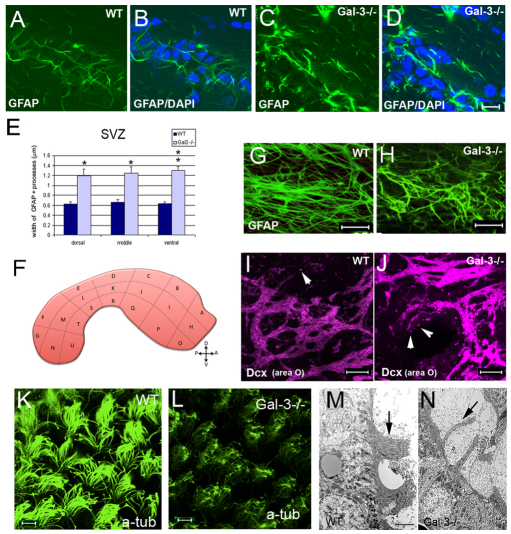
Loss of Gal-3 disrupts SVZ cytoarchitecture. (A,B) The dorsal SVZ contains astrocytes with fine processes exhibiting moderate levels of GFAP immunofluorescence. (C,D) Gal3-null mice had increased levels of GFAP immunofluorescence, thickened processes, and overall distorted astrocytic morphology in the dorsal SVZ. (A–D) Coronal sectons. Photomicrographs in A–D were all taken with the same camera settings. Scale bar: 10 μm. (E) Quantification shows significant increases in the width of GFAP+ processes in the dorsal, middle and ventral SVZ. *P<0.05; **P<0.005. (F) Schematic showing whole-mount dissection of the lateral ventricle wall and the subdivisions used for analysis. (G,H) Whole-mount immunohistochemistry showed that the normal orientation of GFAP+ processes is disrupted in Gal3−/− mice. Scale bar: 20 μm. (I,J) Whole-mount immunohistochemistry shows that more Dcx+ neuroblasts are migrating individually (arrowheads) in Gal3−/− mice compared with the control. Gal3−/− mice also had higher levels of Dcx immunofluorescence than WT in whole mounts. Scale bars: 20 μm. (K,L) Loss of cilia on ependymal cells in Gal3−/− mice is observed using anti-acetylated tubulin antibodies. Scale bar: 20 μm. (M,N) Electron microscopy also shows fewer ependymal cell cilia in Gal3−/− mice. Scale bar: 1 μm. All error bars represent s.e.m.

