Fig. 3.
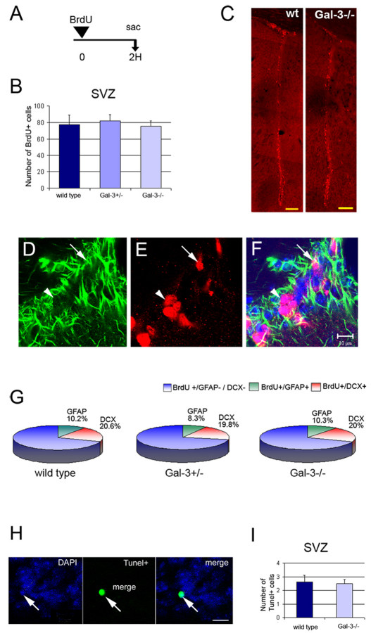
Gal-3 does not affect SVZ proliferation or apoptosis. (A) Short-term BrdU regimen used to assess proliferation. (B,C) The number of BrdU+ cells in the SVZ is not different in Gal-3 mutant mice compared with WT littermates. C shows a low magnification view of BrdU+ cells in the lateral ventricle wall at a mid-striatal A–P level. Scale bars: 250 μm. (D–F) Confocal microscopy of GFAP immunofluorescence (D), BrdU (E), and merge with DAPI (F) from a Gal3−/− mouse. Arrow shows a BrdU+ GFAP+ cell and arrowhead, a BrdU+ GFAP− cell. Scale bar: 10 μm. (G) The percentage of BrdU+ cells that are GFAP+ or Dcx+ is not different in Gal-3 mutant mice. (H,I) The number of Tunel+ cells in the SVZ is not altered in Gal-3 mutants. Scale bar: 5 μm. Error bars represent s.e.m.

