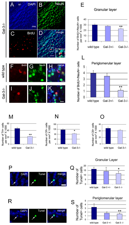
Fig. 5.
Gal3-null mice have fewer new OB neurons. (A–D) Examples of BrdU and NeuN immunoreactivity in the OB granular layer of Gal3−/− mice. Note that most BrdU+ NeuN+ cells are in the deep granular layer near the RMS. (ipl, internal plexiform layer). Inset shows a double-labeled cell (yellow) at higher magnification. Scale bar: 50 μm. (E) The number of BrdU+ NeuN+ cells is significantly decreased in the granular layer of Gal3−/− mice. **P<0.001. (F–H) BrdU+ NeuN+ cells in the periglomerular layer of the OB of a WT mouse. Scale bar: 10 μm. (I–K) BrdU+ NeuN+ cells in the periglomerular layer of the OB of Gal3−/− mice. Scale bar: 10 μm. (L) The number of BrdU+ NeuN+ cells is significantly decreased in the periglomerular layer of Gal3−/− mice. **P<0.001. (M–O) The number of TH+ and CR+, but not CB+, cells decreases significantly in the OB of Gal3−/− mice. *P<0.05, **P<0.005. (P,Q) The number of Tunel+ cells is significantly decreased in the OB granular layer. Arrows indicate a typical Tunel+ cell. Scale bar: 10 μm. *P<0.05. (R,S) The number of Tunel+ cells is significantly decreased in the OB periglomerular layer. Arrows indicate a typical Tunel+ cell. Scale bar: 10 μm. **P<0.005. All error bars represent s.e.m.

