
Fig. 2.
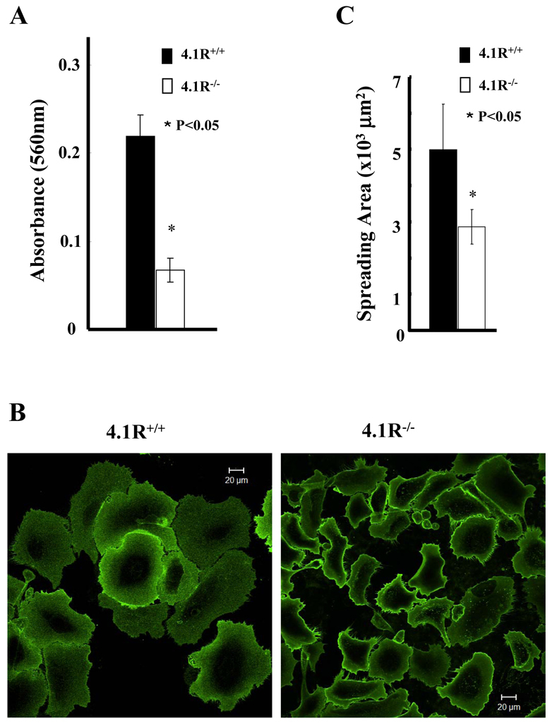
Impaired adhesion and spreading of 4.1R−/− keratinocytes on fibronectin. (A) Cells were plated on fibronectin-coated 96-well plates and incubated for 4 hours. The adherent cells were stained with Crystal Violet and the staining intensity was quantified by spectrophotometry at 560 nm. The results are mean ± s.e.m. of three independent experiments. (B) Cells were plated on fibronectin-coated four-well chambers and allowed to spread for 12 hours. The cells were labeled with Alexa-Fluor-488-conjugated WGA (Invitrogen) and the images were collected using a Zeiss Axiophot wide-field epifluorescence microscope. Scale bars: 20 μm. (C) The mean surface area from 35 individual cells was calculated using LSM 5 Pascal software. The data shown are mean ± s.e.m. of three experiments. One-tailed Student's t-tests were applied to test the statistical significance of the data.